
|Articles|February 1, 2007
Biomarkers aid early detection of joint disease, bone damage
Author(s)Ed Kane, PhD
Bone and articular cartilage turnover in horses occurs both in normal and abnormal states, in healthy and in damaged tissue.
Advertisement
Bone and articular cartilage turnover in horses occurs both in normal and abnormal states, in healthy and in damaged tissue.
In healthy cartilage, a slow constant turnover of the extracellular matrix occurs, and the rate of degradation does not exceed that of replacement. Bone formation and resorption regularly occur to repair skeletal microdamage and to maintain calcium homeostasis. During these processes, metabolites are released in a fairly steady state.
In diseased cartilage, however, various elements are liberated in excessive or unusual amounts, indicating a more severe situation. But there is a benefit:
The ability to quantify degradation and formation products of bone and cartilage matrix elements can help in detecting disease or damage to equine joints, structural changes to articular cartilage, and potential bone damage prior to catastrophic injury.
The biomarkers of interest to clinicians are those of joint disease, osteoarthritis (OA) and bone disease or fracture that result in lameness and pain to horses and trauma to bone and joint tissue, those that are indicators of abnormal skeletal tissue turnover.
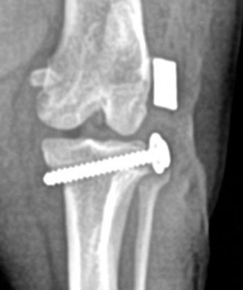
The potential for these markers, used along with radiographs and MRI, is to help equine practitioners detect/diagnose subtle or early damage to tissues, possibly prior to overt clinical signs or obvious lesions seen in x-rays.
During the last several years, researchers have developed assays to help clinicians use these biomarkers as a part of their diagnostic arsenal.
Bone turnover
Bone is composed of mineral, and protein matrix, of which collagen Type I is the main organic constituent, making up about 80-90 percent. The remaining 10-15 percent is of non-collagenous proteins, including osteocalcin, glycoproteins and proteoglycans. The collagen matrix is made sturdy and durable by mineralization with hydroxyapatite crystals.
Within normal bone there is a balance of anabolic and catabolic processes of bone turnover. Bone formation is mediated via the osteoblasts, bone resorption via the osteoclasts, both resulting in release of various molecules. These molecular entities, or biomarkers, are indicative of bone turnover rate. And they can be measured in serum and urine as indicators of the physiological process.
Cartilage turnover
Cartilage turnover is maintained via the chondrocytes that make up 0.15-0.2 percent of the total cartilage volume. The chondrocytes are protected by the extracellular matrix they produce. The primary constituents of the cartilage matrix are Type II collagen and the proteoglycan, aggrecan, which are synthesized and secreted from the chondrocyte. The dry mass of cartilage is about 60 percent collagen Type II, 20 percent proteoglycans. As water is drawn into the matrix, it produces the stiff elastic polymer resistant to impact loading.
With disease, the rate of cartilage degradation exceeds cartilage synthesis, producing a thinner, weaker and less resilient tissue.
Disease, i.e. osteoarthritis (OA) in horses, results from greater degradation and decreased repair of articular cartilage tissue, producing breakdown and erosion of the cartilage surface via extracellular proteineases.
However, early in the disease process, there is some synthesis. Several cartilage protein breakdown products are released into the synovial fluid, blood and urine, which can be measured as disease indicators.
Indirect and direct biomarkers
The disease process involves inflammation of the joints, pain, and loss of flexibility and movement, along with a disturbance of the homeostasis of turnover, producing an imbalance of formation and degradation products.
In OA, there is cartilage breakdown, inflammation of synovial membrane and production of cytokines and enzymes. As the disease proceeds, there is progressive loss of cartilage and subchondral bone change. These changes produce various products which can be measured.
Biomarkers are direct and indirect indicators of abnormal skeletal turnover.
Advertisement
According to Dr. C. Wayne McIlwraith, BVSc, PhD, FRCVS, Dipl. ACVS, Barbara Cox Anthony Chair, director of the Colorado State Equine Orthopedic Research Center (ORC), in equine disease, "the ideal biomarker/s should:
- Detect joint damage at an earlier stage than conventional methods.
- Provide information on disease activity and progressive joint damage.
- Predict future illness and cause of disease.
These biomarkers "can be used to clarify pathobiological processes in the joint, and differentiate diagnostically between affected and non-affected joints, and distinguish the degree of degradation in articular cartilage."
Direct biomarkers originate from cartilage and boney tissue changes, and provide information about their anabolism or catabolism. In cartilage, they are primarily from breakdown products of Type II collagen and proteoglycan fragments. These elements are then released in increased amounts into synovial fluid and thereafter to the blood. In bone, osteoclasts produce breakdown products primarily from Type I collagen. From the anabolic process, osteoblasts and chondrocytes produce proteins that are a part of bone and cartilage formation, surpluses of which can be used as biomarkers of bone and cartilage formation.
Indirect biomarkers are from outside the bone and cartilage tissue, and include proteolytic enzymes, pro-inflammatory cytokines and other molecules. These indirect biomarkers include matrix metalloproteinases, IL-1, tumor necrosis factor (TNF?), hyaluronic acid and C-reactive protein. These indirect products are produced in reaction to stress or damage to the joint and/or bone.
While direct biomarkers have been shown to be of value for clinicians as indicative of disease, indirect biomarkers, that vary more subtly, have been shown to be of value to research situations, but not as yet practical as diagnostic tools.
Direct biomarkers of cartilage metabolism
Anabolic biomarkers include carboxypeptide of Type II collagen (CPII) and CS-846, a marker of chondroitin sulfate (CS) synthesis.
CPII is a product of Type II collagen synthesis, and has been seen in elevated amounts in blood serum of horses with osteochondral fragmentation, since, according to McIlwraith, "it appears that increased synthesis of Type II collagen is an early reaction to osteochondral fragmentation." In human OA patients, CPII is increased several fold in synovial fluid, but decreased in blood serum.
CS-846 is a useful marker of aggrecan synthesis. In a study in horses with osteochondral damage of the knee (carpus), an epitope, (CS-846) was found to be significantly higher in joints with damage and in serum of those horses. A majority (79 percent) of horses with osteochondral fragmentation had significantly higher amounts of serum CPII and CS-846. In human OA patients, it was found that circulating levels of CS-846, are highest in patients with the longest time period of disease, and in those with greatest cartilage loss.
With cartilage breakdown (catabolism), antibodies have been developed to identify Type II collagen fragments in horses with OA and OCD. Significant increases in degraded Type II collagen have been measured in synovial fluid and serum samples from horses with articular damage. In a study of OC in foals, it was found that there were higher amounts of COL2-3/4C and 234 CEQ assays detecting collagenase-cleaved Types I and II collagen fragments, and Type II collagen fragments, respectively.
Also with cartilage degradation, significantly higher levels of glycosaminoglycans (GAGs) were found in synovial fluid samples from horses with OCD and traumatic arthritis.
In another study, sulfated GAG concentration was significantly higher in horses with severe to acute, moderate to chronic, and severe to chronic joint changes, as well as in those with arthroscopic lesions. Horses with experimentally produced early OA have also been shown to have higher serum levels of GAGs.
In human OA patients (knee), higher serum levels of hyaluronan, a high-molecular-weight GAG, were found and in other arthritis patients, elevated serum hyaluronan predicted more pronounced progression of the disease.
In human OA and RA patients, YKL-40, a cartilage glycoprotein (found in low concentration in normal cartilage) has been detected in increased amounts in synovial fluid and serum from patients with "active" RA, and in those with "late-stage" knee OA.
Another promising cartilage breakdown product, glucosyl-galactosyl-pyridinoline, involved in cartilage cross-linking, has been shown in human clinical studies in patients with RA or OA to be released and found in increased amounts in urine.
Though possibly not as useful in predicting joint damage, there is some evidence for measurement of keratan sulfate (KS) in horses with moderate to chronic and severe to chronic joint disease. Keratan sulfate is commonly found in the proteoglycans molecules of aggrecan. A study looking at the value of KS levels in horses with osteochondral fragmentation found that it had little or no value.
Another biomarker of cartilage breakdown is cartilage oligomeric matrix protein (COMP), an abundant noncollagenous protein constituent of articular cartilage.
According to McIlwraith, "initial work assessing a COMP assay in serum and synovial fluid from horses with joint disease is promising."
In another study, "the levels of COMP and aggrecan were reported in synovial fluid from carpal joints of acutely lame Standardbred and Thoroughbred racehorses with macroscopic lesions of articular cartilage (OA), osteochondral fractures, and ligament tears found at arthroscopy."
There was increased concentration of COMP in synovial fluid of Thoroughbreds with osteochondral fractures, but not from joints with OA. It was concluded that "an elevated synovial fluid COMP concentration could be a useful marker for diagnosis and monitoring of equine middle carpal joint fractures in Thoroughbreds."
Unlike in horses, increased synovial fluid and serum levels of COMP were found in human OA patients, and an increased serum COMP with the disease activity of OA, as well as a correlation with the radiographic progression of the joint disease, as well as with indicators such as pain, joint stiffness and physical function.
It has been shown that high levels of aggrecanase, the principal protein responsible for aggrecan degradation in articular cartilage, are found in synovial fluid of diseased equine joints.
Urinary levels of CTX-II (a degradation marker) have been associated with disease activity of human OA, and have been shown to be associated with radiographic progression of disease.
Direct biomarkers of bone metabolism
McIlwraith states that "biomarkers of bone degradation and synthesis can be valuable in determining the stage of subchondral and epiphyseal bone disease, predicting fracture, and monitoring the role of bone in joint disease." Most of the biomarkers of bone turnover are of Type I collagen, the largest amount of protein of the bone matrix, and also assays of non-collagenous bone proteins.
Carboxy and amino terminal propeptides (PICP and PINP), cleaved during bone synthesis, may potentially be used as markers of bone formation, though little evidence so far has been found for their association with equine bone disease/damage.
Osteocalcin, a small protein associated with bone formation and turnover, has been studied to see if it might be of value to identify horses with bone disease, though, up to now, there has been poor evidence to show its value in horses. There is, however, some data in humans with OA.
Bone-specific alkaline phosphatase (BAP), shown at high levels in bone-forming osteoblasts, plays an important role in bone formation. Though onset of race training has shown an increase in BAP, little has been seen so far for its use in diagnosing bone disease/damage. It would be logical for increased amounts to be found in horses in training, in that, for such horses, microdamage and repair would be a normal consequence. It might be of value for predicting outright bone fracture and repair, but so far there are no data.
Type I collagen non-helical telopeptide (ICTP), a cleaved fragment of bone turnover, has been measured in horses in relation to age, exercise and breed differences, but it has not been shown to be of value for diagnosis of bone disease.
Type I collagen C-telopeptides (CTX), though showing possible promise in evaluating human OA, has shown little value regarding equine OA.
Bone sialoprotein (BSP) found to be elevated in human OA patients, and increased on immunohistochemical examination of tissue in horses with degenerative changes of the bone and cartilage, is thought to have "the potential as a biomarker for changes in bone metabolism in subchondral bone." It is suggested by McIlwraith that "further studies should focus on the presence of BSP in the synovial fluid and serum from racehorses with different types and aetiology of OA lesions."
Future value of biomarkers
Recently, Drs. McIlwraith and David Frisbie at the ORC, in collaboration with DVMs Vince Baker, Jeff Blea and Rick Arthur in California, have shown certain markers to be increased prior to injury in a cohort study of Thoroughbred racehorses.
While various biomarkers discussed show value and some show promise in diagnosing present and pending bone and joint damage/disease, there may be limitations to their uses.
An animal may have local disease, but the biomarkers found in serum and urine are an indication of the total metabolic activity of the cartilage/bone, therefore originating from all the joints/bones in the body, not necessarily limited to reflection of disease in a few or a specific joint/bone.
Biomarkers are therefore systemic by their nature.
That is possibly why some do not always reflect the clinical picture affecting one or a few joints/bones, and their use for this purpose may be limited in some cases.
In other cases, it has been shown that changes in biomarkers, reflecting disease in one or a few joints/bones will show significant changes, can be measured and be of diagnostic value.
Newsletter
From exam room tips to practice management insights, get trusted veterinary news delivered straight to your inbox—subscribe to dvm360.
Advertisement
Related Content
Advertisement

Advertisement
Advertisement
