Approach to otitis, client education leads to success (Proceedings)
Treatment of otitis externa is dependent on identifying and controlling the predisposing factors, primary and secondary causes and perpetuating factors whenever possible. Inadequate treatment and reversal of the progressive pathologic responses, tympanic membrane alterations, and otitis media often leads to treatment failures or recurrences.
Goals
Treatment of otitis externa is dependent on identifying and controlling the predisposing factors, primary and secondary causes and perpetuating factors whenever possible. Inadequate treatment and reversal of the progressive pathologic responses, tympanic membrane alterations, and otitis media often leads to treatment failures or recurrences. Even though externally and to the client the ears seem better, adequate treatment can only be determined by otoscopic examination and follow up cytological examination of material from the ear canal. The treatment of otitis externa often requires a complete plan that will involve the use of multiple components. It is essential for these components to be successful that there is good client compliance. In general achieving compliance is greatly facilitated when the client understands the problems and corresponding goals. It is critical that follow up examinations are performed and clients or the clinic effectively keep the ear canal clean from excessive build up of debris, microbes or exudates. The client must be able to properly apply topical medications or an alternative form of therapy is required.
PSPP System© For Otitis Externa
Whenever a case of otitis, especially chronic otitis, presents then the PSPP System (see table 1 and www.animaldermatology.com) should be used to help establish what is really happening in the ear and educate the client.

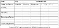

Table 1 from PSPP System©
This is done by entering a list of potential components of their pets ear disease and explaining them to the owners in a visual method, with simple drawings. The drawings help educate the owner about normal ear anatomy and physiology as well as perpetuating factors that may occur. Each patient has their information entered into the table. Tests and treatments for each component of the pets problem are listed and categorized prognostically, cured, requires long term management to achieve control or will require life long treatment. This simple system greatly facilitates educating clients about their pets problem. It gives the client reasonable expectations about the prognosis and what will be involved in "fixing" their pet. This results in improved client compliance and minimizes client frustration and searching for a "better" veterinarian.
Why Clean Ears
Thorough cleaning of the ear canals is extremely important for the effective management of otitis externa. It is often the most important step and if one treatment only was allowed to manage ear cases the it would definitely be cleaning as not single topical product is as effective. Different types of debris may occur in ears and there presence can impact otitis and the response to therapy. In normal ears little debris is present and it is composed of keratinocytes, cerumen and some microorganisms.
In otitis cases there are multiple reasons why it is important to clean out excessive or abnormal debris. The debris can hinder the penetration of topical agents to the affected tissue requiring treatment, and large deposits may prevent medication from reaching the deeper parts of the horizontal canal, tympanum or middle ear when the tympanum has been compromised. This leads to the number one rule of topical therapy, the ingredient must reach the site to be treated. The debris can protect microbes that are rolled in or attached to keratinocytes and covered with a protective lipid layer. These organisms may survive therapy and then infect the ear again. Larger clumps of debris may remain in folds, middle ears and false middle ear cavities where they sequester organisms. Retained debris may also contain pro inflammatory material such as microbial byproducts and toxins as well as mediators released from inflammatory cells that may be present. Purulent exudates interfere with and bind to antibiotics such as polymyxin and aminoglycosides. Complete otoscopic examination with visualization of canal and tympanum pathology will often require cleaning. In some cases the use of a stronger topical agent or overuse of antibiotics can be prevented by regular intervals of ear cleaning, especially if disinfectants are included in the cleaning process. When progressive pathologic changes result in abnormal epithelial migration or folding in the canal lumen then cleaning will be required until this normal physiologic function returns.
Cleaning of the ear canals is extremely important for the effective management of chronic otitis externa. Cleaning the ear canal is a lot like cleaning a skin fold lesion. With chronicity the lining of the ear canal is thrown into proliferative folds. Management of fold dermatitis requires surgical removal or keeping the skin folds clean. Without surgery successful control often requires keeping the skin folds clean indefinitely or until the ear canal normalizes and the folds are resolved. In chronic cases with otitis media this also includes cleaning the bulla or false middle ear.
Cleaning Methods
Cleaning techniques vary considerable in ease of use and efficacy. There are several techniques commonly utilized with some also simple enough for clients to do at home however deep ear cleaning and cleaning of the middle ear requires retrograde flushing[.
Home Cleaning and Use of Disinfectants
Teaching the clients to clean either with home cleansers that do not require rinsing or a bulb syringe is an important aspect to controlling atopic otitis cases. Cleaning is helpful for several reasons. Debris and microbes are removed thus decreasing overgrowth and the environment favorable to overgrowth. Allergens that are present on the skin surface or trapped within the cerumen and debris on the pinna and in the ear canal are also removed or at least the allergen load is decreased. In severely inflamed, painful ears home cleaning should be delayed as many dogs will not tolerate these procedures until after the inflammation and pain is resolved. Therefore home cleaning is usually recommended following the initial therapy. There are two main ways for clients to clean ears at home.
Ear wash or rinse is the most common method used at home. This technique has the client instill a mild cleanser into the ear until the canal has been filled to the external orifice as well as apply the cleanser to the external orifice and concave pinna. This tends to be a little messy and should be done outside or in a bathroom or even shower stall. There are two important aspects to this technique. First since the cleanser may not be rinsed out is the use of an effective cleanser that is safe to leave in an ear. My favorite cleansers for home use are Douxo® Micellar Cleanser, Sogeval which is preferred by most clients and Cerumene™, Vetoquinol. The second important aspect to this technique in dogs with significant horizontal canal build up is adequate massage of the auricular and annular cartilage. To effectively massage the annular cartilage the client must be educated about the location and need for deep digital palpation. Following several minutes of massage the material is allowed to be shaken out and then the external orifice and concave pinna is wiped clean with tissue or cotton balls. Do not allow excessive use of cotton tipped applicators down the ear canal as these commonly push debris deeper into the ear canal. In general this rinse technique is not as effective as bulb syringing due to the inability to massage with fluid the last portion of the horizontal ear canal which is contained within the boney external acoustic meatus.
Bulb syringing is generally more effective in cleaning the horizontal canal and gets the ear canal cleaner than the wash/rinse technique. It is somewhat more labor intensive, messy and requires that clients are adequately trained and the patient will tolerate the procedure. This technique is not usually used home rinses have been shown to be ineffective. In general the tympanum should be intact if cerumenolytics are going to be used because the client may not be able to adequately rinse away all residual drugs and repetitive application could be dangerous especially if not being rinsed out. Clients should be instructed about the proper pressure and speed to squeeze the bulb syringe. This is best determined by initially practicing with air and learning how hard and fast they can squeeze without hearing the air coming out of the bulb syringe. Only lukewarm or body temperature fluids should be used for flushing. The nipple of the bulb syringe should be placed loosely in the external orifice of the ear canal so that once flushing is started they rapidly see the flush solution exiting the ear canal. Allowing adequate back flow helps prevent excessive pressure building up against the tympanum. Flushing is continued with fresh solution until no debris is seen coming from the ear canal. They should not use the bulb syringe to aspirate fluid out of the ear as the inside of the syringe may become contaminated with pathogenic organisms. Following flushing with a bulb syringe a cleanser disinfectant or disinfectant dryer solution is used to help dry the ear canal and remove residual rinse water.
Disinfectants may be utilized as an ear rinse or following the home cleaning by either method. Disinfectants have the advantage of not inducing resistance and are often less expensive than antibiotics. Ingredients often used for disinfectant properties are isopropyl alcohol and one or more of the following: acetic acid, boric acid; benzoic acid; lactic acid, malic acid, salicylic acid, silicone dioxide, sulfur. Just lowering the pH of the ear also is antimicrobial and some products such as those with citric acid and sodium citrate function by this mechanism. Combination of 2% boric and 2% acetic acid is available commercially. (Malacetic, Dermapet®) This type of product has been shown to have some efficacy in controlling secondary infections though one study showed 2% acetic/ 2% boric acid product poor as a prophylactic agent for prevention of Malassezia otitis. A combination of tris edta and 0.15% chlorhexidine was shown to have a synergistic effect and has been very effective in Pseudomonas cases. This combination is available in Europe (Otodine, ICF) and in the US (TrizEDTA™ Plus, Dermapet®) but is not approved for otic use in the US. Phytosphingosine (Douxo® Seborrhea spot on) besides being incorporated into the lipid barrier and having some possible anti inflammatory effects in the ear may also have antimicrobial effects. Anecdotal reports of benefit in otitis have led to a blinded study that is currently being done in otitis cases.
Setting Up and Selling Follow Up
The first step in this process is developing the recheck plans and getting the client to understand their importance and follow through. Repetitive examinations are very important, especially in cases where the dog is apparently improving. Though clinically the odor, head shaking and discomfort may be gone the ear may still be building up debris and not staying cleaned, have proliferative changes, or the tympanum may not have returned. These changes may eventually lead to another infection or acute flare up of otitis. The return to a healthy ear canal can only be determined with otoscopic and cytologic examination of the ear canal. Follow up examinations are also important to determine if cleaning is being done effectively and when normal self cleaning returns. Scheduling the follow up examination is critical and has to be done differently to answer the preceding questions. To determine if home cleaning is effective then the examination should be done within 24 hours of the cleaning procedure. To determine if the interval is to long between cleanings or that self cleaning may have returned the examination needs to occur when the ear has not been cleaned for at least the interval between current cleanings or longer. For example a dog has been doing well and the client has been cleaning at home every week. The next examination may be scheduled when they have not cleaned the ear for 10- 14 days so that it can be determined if the ear is staying relatively clean between cleaning procedures. Clients need to understand there are different types of follow up for chronic ear cases and that multiple visits will be required.
References
Nuttall, T. and L.K. Cole, Ear cleaning: the UK and US perspective. Vet Dermatol, 2004. 15(2): p. 127-36.
Griffin, C., Otitis Externa and Media, in Current Veterinary Dermatology, The Science and Art of Therapeutics, C. Griffin, K. Kwochka, and J.M. MacDonald, Editors. 1993, Mosby Year Book: St Louis.
Ghibaudo, G., L. Cornegliani, and P. Martino, Evaluation of the in vivo effects of Tris-EDTA and chlorhexidine digluconate 0.15% solution in chronic bacterial otitis externa: 11 cases. Vet Dermatol, 2004. 15(s): p. 65.


