Anatomy of the thoracic viscera (Proceedings)
The respiratory system extends from the tip of the nose to the diaphragm.
General comments:
• The respiratory system extends from the tip of the nose to the diaphragm.
• Take good quality radiographs.
• Take as many radiographs as needed to make the diagnosis or to determine that other diagnostic methods are needed.
• If the pet is anesthetized, it takes less than 60 seconds for atelectasis to occur; use forced inspiration/mechanical inspiration to expand the lungs while taking the radiograph.
• Take sequential radiographs to monitor a disease process and response to therapy.
• Have the radiographs reviewed by the radiologist if you are not sure what is happening.
• CT imaging is a more "robust" technology to assess the lungs especially for "mass" lesions.
• Ultrasound is a more "robust" technology to assess the cardiac silhouette and heart.
• Intervention may be required to obtain a definitive diagnosis.
• Some animals never get completely better and others die despite your best efforts
The Airways and the Lungs
The number of radiographic projections required to define a lesion will depend on the extent and location of the lesion. The common views are:
A. Ventrodorsal projection:
• The sternum is towards the x-ray tube; the vertebrae are against the cassette or on the table.
• This view is obtained to evaluate the cranial mediastinum, ventral contour of the heart and ventral lung region.
B. Dorsoventral projection:
• The vertebrae are towards the x-ray tube; the sternebrae are against the cassette or on the table.
• This view is obtained to evaluate the caudal mediastinum, overall contour of the heart, the aorta and the dorsal lung region.
• In both of these projections, the x-ray beam is centered on the fifth thoracic vertebra; the vertebrae and sternebrae are superimposed; the elbows are adducted, and the skin folds are moved laterally.
C. Right lateral projection:
• The left lateral surface of the torso is toward the x-ray tube; the right lateral surface of the torso is against the cassette.
• This view is obtained to evaluate the left lung field, mediastinal structures, left side of the diaphragm, esophagus and pericardial sac.
D. Left lateral projection:
• The right lateral surface of the torso is toward the x-ray tube; the left lateral surface of the torso is against the cassette.
• This view is obtained to evaluate the right lung field, mediastinal structures, right side of the diaphragm and pericardial sac.
• It is also obtained to evaluate for a hiatal hernia after barium administration.
• In both lateral projections the beam is centered on the fifth intercostal space; the sternum is lifted slightly away from the table; the forelimbs are pulled forward, and the skin folds are moved dorsally and/or ventrally.
E. Oblique projection:
• This view is used to remove superimposition
• This view is used to "highlight" an area of interest by removing superimposition of tissues.
Radiographs of the thorax are usually obtained during inspiration unless you suspect pneumothorax or free pleural fluid. Free gas or fluid is easier to define when the animal is in expiration.
• The lung, when viewed in expiration, is often mistakenly diagnosed as being pathologic. When the animal is in expiration, there is less gas in the lung, and the lung becomes more opaque. This physiologic change results in increased lung opacity that mimics many disease processes that also cause the lung to become more opaque.
• Air/gas is the natural contrast agent of the lung; take advantage of this contrast agent by radiographing the animal in inspiration when you wish to evaluate lung parenchyma. The pericardial sac, mediastinal structures and the diaphragm.
• When the animal is in expiration, the pulmonary vessels are closer together, the diaphragm overlaps the heart; the cranial lung lobes do not extend to the thoracic inlet, and the pericardial sac appears larger.
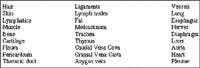
Anatomical structures:
The x-rays pass through many structures when the thorax is radiographed, and all of these structures can have different presentations depending on their configuration. These structures are:

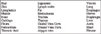
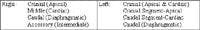
Lung lobes:


Overview of the airway:
• Nostrils (Nares)
• Nasal Cavity
• Nasopharynx
• Hyoid bones:
o Stylohyoid
o Basihyoid
o thyrohyoid
o epihyoid
o ceratohyoid
• Larynx
• Trachea
• Bronchi
• Bronchioles
• Laryngeal ventricles
• Epiglottis
• Cricoid cartilage
• Thyroid cartilage
• Arytenold cartilage
• Cuneiform process
o corniculate process
o Sesamoid cartilage
• Division of Airways –
o Trachea, Principal bronchi (primary, main stem)
o Lobar (secondary)
o Segmental (tertiary)
o Sub-segmental
o Respiratory Bronchioles
o Alveolar ducts
o Alveolar Sacs
o Alveoli.
Cartilaginous elements no longer present when bronchioles less than 1mm in diameter.