Advancing laminitis research - a collaborative effort
Kennett Square, Pa. - Research into the causes and treatment laminitis, along with wide-ranging collaboration are the driving forces behind the newly established Laminitis Institute at the University of Pennsylvania.
Kennett Square, Pa. — Research into the causes and treatment of laminitis, along with wide-ranging collaboration, are the driving forces behind the newly established Laminitis Institute at the University of Pennsylvania School of Veterinary Medicine's New Bolton Center.
"We want people to know that the Laminitis Institute is up and running, and that we have a mission and vision for the future — we are committed to making sure the next generation of researchers/clinicians see laminitis as an important area for investigation and even solving the puzzle," said Dr. James Orsini, the institute's director, in a recent statement through the university.


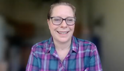
Photo 1: From left, Dr. Christopher Pollitt, director of research; Dr. Hannah Galantino-Homer, lead research investigator and Dr. James A. Orsini, director of the Laminitis Institute at Penn Vet. (Photos: University of Pennsylvania New Bolton Center)
The institute was established primarily with a gift from philanthropists Marianne and John K. Castle, along with the Barbaro Fund, launched after the Thoroughbred's death in 2007 following a bout with laminitis.
Christopher Pollitt, BVSc, PhD, a world-renowned laminitis expert, was recently named research director for the institute, joining Dr. Hannah Galantino-Homer, the senior lead investigator. Pollitt will coordinate the work of researchers worldwide, including his own Laminitis Research Unit at the University of Queensland in Australia.


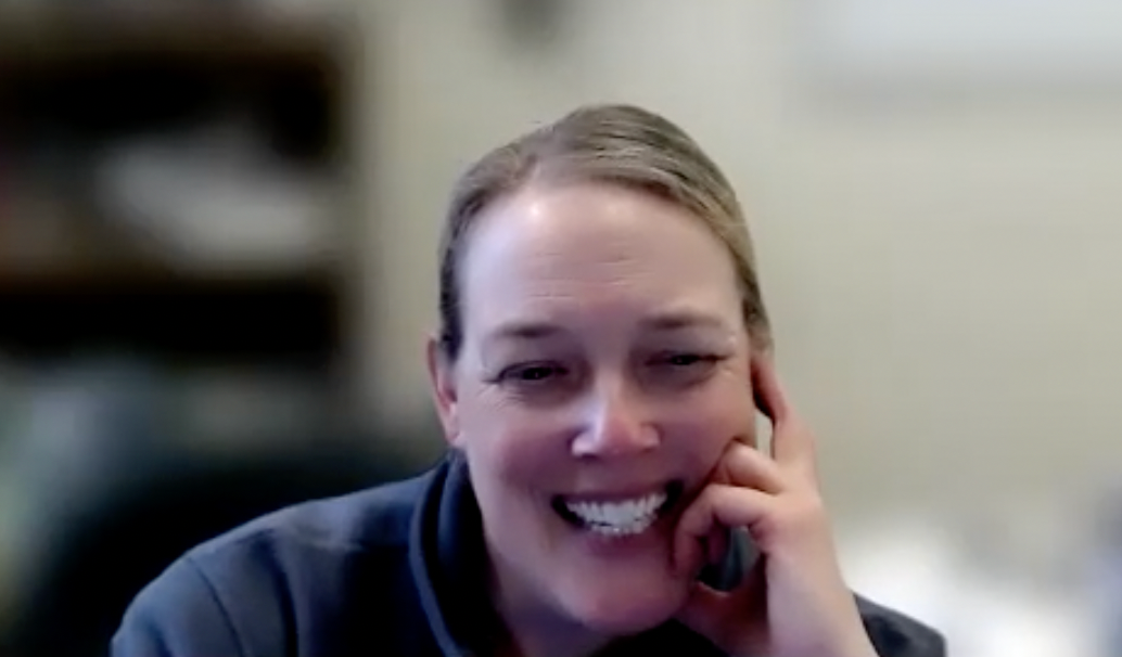
Photo 2: Investigation into the pathogenesis of the disease is a main focus of Penn Vet's laminitis researchers.
Already he is advancing the institute's first major initiative — creating a tissue bank at New Bolton Center. Tissue and blood samples from horses with either experimentally induced or naturally acquired laminitis, along with data, are being collected from researchers throughout the United States and worldwide, with the institute serving as a repository for all of them to access.
Research goals
"Our goal is to take a three-pronged approach to answer some very basic and general questions," says Galantino-Homer, VMD, PhD, Dipl. ACT, the lead investigator. "Gleaning from the human and veterinary literature, and proving through our experiments which pathways apply and which don't, we hope to find some answers."
The three research components are tissue banking, investigation into the pathogenesis of laminitis and developing an in vitro lab model of laminitis.
The tissue bank is mainly to provide tools for researchers. "We have an assortment of tissues from laminitic and non-laminitic horses," says Galantino-Homer.


The laminitis institute 4-point strategic plan
Included are serum and tissue samples from several Cushing's horses, some with laminitis and some without. "We have all the information that goes with those samples — endocrine studies for the Cushing's horses, laminar histopathology, what research has been done with samples so far, everything centralized because it's not always easy to get that material to work with," says Galantino-Homer.
The Bernice Barbour Foundation recently provided a three-year grant to develop a laminitis discovery database, which will include the tissue and serum bank, the information associated with the samples and a database of information on antibodies that can be used for equine studies.
The Grayson-Jockey Club Research Foundation, working with Pollitt, is using tissues from oligofructose and hyper-insulinemic clamps, two of Pollit's models of in vivo laminitis.
The first of Pollitt's two models of inducing horses with laminitis is feeding oligofructose, a commercially available, natural carbohydrate, similar to the carbohydrate derived from many temperate grasses.
"Once you dose the oligofructose into the horse, it changes the microflora of the hindgut dramatically and very rapidly," says Pollitt. This produces an overgrowth of Streptococcus equinus complex, a gram-positive bloom in the hindgut. This makes a horse moderately ill and initiates the beginning stages of the disease at the basement membrane of the lamellae in the hoof.


Figure 1: Normal anatomy of the distal phalanx/coffin bone and related structures in the equine foot. (Figures: Molly Higgins/University of Pennsylvania School of Veterinary Medicine)
"The (institute's) plan is to repeat that model, but at much earlier time points," Pollitt explains. "We want to track back to the very earliest molecular events that are occurring to give us an insight into how soon this bloom of microbes in the hindgut affects the lamellae of the feet."
Institute researchers have good evidence that as early as 12 hours post-dosing, significant changes occur in the lamellar basement membrane. They will use the in vivo model, collecting samples of tissue from the basement membrane as early as six hours, 12, 18 and 24 hours post-dosing.
There is no need to produce end-stage laminitis per se; the horses barely notice the effect of the induction and effects of the oligofructose in their hindgut.
"At this time we know plenty about the eventual disease. Our goal now is to study the very early induction phases, and ethically this is preferable," Pollitt says.
The samples will going to a leading equine genomics expert, Bhanu Chowdhary, BVSc & AH, MVSc, PhD, professor in the Department of Veterinary Integrative Biosciences at Texas A&M University, who will perform a genomic analysis with the full equine genome micro array. "This is a major advantage of the laminitis institute," says Pollitt. "We have a researcher consortium around the world, from Australia, to Texas, to Penn Vet."


Figure 2: Rotation of the distal phalanx/coffin bone due to the compromised bond between the epidermal and dermal lamellæ tissue along the front of the foot.
Galantino-Homer's lab will be doing a proteomic analysis of the blood and tissues from the horses at the early time points to note the early events.
Pollitt's other model is a hyperinsulinemic model, working with Standardbred horses. Insulin is dramatically laminitogenic. Researchers can induce laminitis in Standardbred horses as soon as 48 hours by making them hyperinsulinemic while keeping their glucose levels normal, using a euglycemic-hyperinsulinemic clamp extended to 48 hours. "It's quite difficult to do," says Pollitt, "but it provides quite dramatic results."
The Grayson research involves gene expression and proteomics (study of component proteins) to note expression differences between the control and the induced horses to try to fill some gaps in the pathogenesis.


Figure 3: Sinking of the distal phalanx caused by complete loss of the bond between the epidermal and dermal lamellæ tissue.
"In that effort we are working with our epithelial stem-cell colleagues on ways to culture the basal cells, the epidermal lamellar cells that are right at the interface with the basement membrane," says Galantino-Homer.
"We assume that those cells are going to be at ground zero, regardless of the initiating cause of laminitis."
Whether it's the case of these cells proliferating when they're not supposed to be, releasing proteases that break down the basement membrane, being affected by inflammation or responding to proposed laminitis trigger factors, those are the cells of interest.
If funding allows, the institute would like to try to develop biomarkers to help identify horses at risk for laminitis.
Integrated research
The laminitis institute's work is integrated into various schools at Penn Vet, including the animal biology and pathology departments and the University of Pennsylvania School of Medicine. "I feel like this project is at the heart of the school," says Galantino-Homer. "Everybody wants to be involved. It's been great to have so much interest and support from such a wide cross-section of the Penn community."
"The tissue interface where laminitis occurs is structurally very complex," notes Julie Engiles, VMD, Dipl. ACVP, a veterinary pathologist and another member of the group. The hard (insensitive) portion of the hoof wall is integrated with the sensitive laminae, which are epidermal tissues. Beneath that lies a complex vascular network supported by a dense (connective) tissue that is tightly adhered to the pedal bone.
The destruction of the sensitive laminae that occurs in laminitis will by nature affect the other elements.
At this point, it is not known if, or how these various integrated tissues (epidermis, connective tissue, nerves, vasculature and bone) affect or exacerbate disease progression, clinical symptoms (e.g., pain/lameness) or tissue regeneration/repair.
Often these tissues are not evaluated on routine pathologic examinations. Even in cases of laminitis, many times only gross postmortem evaluation of these tissues is performed. Therefore, work is needed on systematic histomorphologic characterization of laminitis in naturally occurring disease.
In addition, the complex tissue interface of these tissues implies complex molecular pathways to maintain normal physiologic function, another area that has not been fully evaluated in either normal or laminitic animals (Dr. Galantino-Homer's focus).
Engiles' research focuses on the systematic histopathologic characterization of the pedal-bone-hoof interface in laminitic animals, and the correlation of these changes with the changes that occur at the molecular level.
"Potentially we'll end up with specific biomarkers or measurable parameters of the disease process for early detection of laminitis, or perhaps for future treatments," Engiles says.
A serious move toward sharing laminitis research came from a 2004 meeting in Louisville, Ky., convened by the American Association of Equine Practitioners (AAEP) and organized by Rustin Moore, DVM, PhD, Dipl. ACVS, who heads the Department of Veterinary Clinical Sciences at The Ohio State University Veterinary Teaching Hospital and who serves on the laminitis institute's scientific advisory committee.
"One of the goals from that meeting," Moore says, "was to promote increased collaboration and more widespread sharing of information and (tissue and blood) samples ... so that we could learn together. If we're going to be successful, this needs to be a collaborative effort."
Besides Penn Vet, active laminitis research programs are ongoing at the University of Georgia, Louisiana State University, University of Missouri, University of Tennessee, The Ohio State University and other schools worldwide.
Ohio State's laminitis research program, headed by James Belknap, DVM, PhD, Dipl. ACVS, pointed to the presence and role of inflammation, leading to a paradigm shift in the way researchers now approach laminitis worldwide.
His studies have recently involved cutting-edge cellular and molecular techniques to introduce the laminitis research and clinical world to the concept that many similar events occur in laminar failure in laminitis as occur in organ failure in human sepsis patients.
These include endothelial activation, activation and infiltration of leukocytes and the involvement of a host of different inflammatory signaling pathways.