Acute spinal cord injury – The first hour can make the difference (Proceedings)
Spinal trauma occurs from external or internal causes.
Principals of acute spinal cord injury
Primary injury
Spinal trauma occurs from external or internal causes: external – automobile-related injury, falls, falling objects, and projectile missile damage; internal – intervertebral disc disease, pathologic fractures, vascular. Mechanisms of primary injury include compression, shear, laceration, bending and distraction. The thoracolumbar region is the most commonly injured are in the dog and cat. Fractures occur between T11 and L6 in 50% to 60% of patients after blunt trauma. Fractures in the thoracic spine may have little displacement because of the protection provided by the ribs, ligamentous support and epaxial musculature. Fracture and luxation of the thoracolumbar spine are often associated with other systemic injuries: pneumothorax, pulmonary contusions, orthopaedic injuries, urogenital injuries, and diaphragmatic hernia. Approximately 20% of patients with thoracolumbar fractures have a second spinal column fracture-luxation. The amount of neural tissue injury is related to the rapidity and severity of insult, and the amount and duration of compression.
Secondary injury
Primary mechanical injury to the neural tissue can subsequently lead to secondary biochemical injury (secondary injury theory). Acute spinal cord injury causes both systemic and local vascular abnormalities. Primary injury ultimately results in a progressive decrease in perfusion and necrosis of the injured area of the spinal cord. Secondary metabolic injury results from ischemia include abnormal release of excitatory neurotransmitters, abnormal accumulation of intracellular calcium, and activation of membrane phospholipases and production of free radicals. Vascular mechanisms include – ischemia, impaired autoregulation, neurogenic shock, hemorrhage, microcirculatory disruption, vasospasm, and thrombosis. Ionic derangements include – increase intracellular calcium and sodium and increased extracellular potassium. Accumulation of neurotransmitters includes – serotonin, catecholamines, and extracellular glutamate. The severity of the pathologic changes and the degree of recovery are related to the duration of acute compression as demonstrated in studies of animal models. New treatments are aimed at those limiting the secondary injury and those that promote regeneration of the injured cord (oscillating field stimulators, polyethylene glycol, 4-aminopyridine).
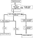
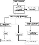
Patient assessment
Initial evaluation
If a spinal fracture is suspected, the patient should be secured and immobilized to a back board to prevent further injury. Additional restraint or sedation may be necessary if the animal is struggling. The animal should be evaluated in the position that it is presented, usually in lateral or sternal recumbency. Spinal injury frequently occurs in association with multiple organ trauma. Basic principles for management of spinal cord injury (SCI) are applied to ensure stabilization of the patient's physical status. Upon admission, the patient is stabilized by assessing airway patency, breathing and circulatory status. Spontaneous respiration and evidence of respiratory compromised are closely monitored in patients with cervical SCI. Blood gas monitoring will assess severity of hypoventilation determined by measuring PaCO2 (35-45 mm Hg). Oxygen supplementation will assist to maintain PaO2 (> 90 mm Hg). Trauma may predispose to primary lung disease associated with pneumonia, atelectasis, hemorrhage or edema.
Fluid therapy
General medical management of patients with acute SCI should include restoring a normal blood pressure, enhancing intravascular volume and avoiding hyperthermia in an effort to enhance spinal cord perfusion. Hypotension will often follow spinal cord injury due to multiple factors: interruption to first order neurons of the sympathetics resulting in unopposed parasympathetic influence and bradycardia; spinal shock causing loss of muscle tone and secondary hypovolemia from venous pooling; and blood loss from other associated injuries. Integrity of the circulatory system is monitored with MABP and electrocardiogram.
Medical management specific to spinal cord injury
The pathophysiology of acute SCI has led to relevant neuroprotective approaches to attenuate the effects of secondary injury. Neuroprotective effects of methylprednisolone sodium succinate (MPSS) include inhibition of lipid peroxidation, calcium influx, ischemia, axonal dieback, cytoskeletal degradation, and anti-inflammatory effects. Methylprednisolone sodium succinate (MPSS) administration protocol is 30 mg/kg IV slow bolus; two hours after the first dose administer 15 mg/kg IV; 4 hours after the second dose administer 15 mg/kg; consider CRI 5.4 mg/kg/h for 24 hours. If more than 8 hours after injury corticosteroids should be avoided. Therapeutic efficacy has been questioned in humans and studies report an increased rate of sepsis and pneumonia. No scientific randomized controlled clinical trials exist with use of MPSS in animals. Complications of corticosteroid therapy in dogs include diarrhea, melena, vomiting, hematochezia, hematemesis, and anorexia.
Neurologic assessment
Results of the neurologic examination are used to determine neuroanatomic localization and severity of the spinal cord injury. It is important to perform the neurologic examination with care to prevent further injury and displacement of the spine. Cranial nerves, spinal reflexes, spinal palpation for hyperesthesia, cutaneous trunci reflex, and assessment for deep pain perception can be performed in lateral recumbency. Presence or absence of deep pain perception is the most important prognostic indicator. Animals that have no deep pain for > 48 hours have a grave prognosis. Dogs with suspected intervertebral disk disease and absent deep nociception for < 24 hours have a 60% chance of regaining ambulatory status. Animals presenting with a spinal fracture and absent deep nociception, the prognosis for walking is hopeless.
Neuroanatomic localization for spinal cord lesions is determined based on presence or absence of spinal reflexes. A C1 through C5 lesion results in tetraparesis/plegia or ipsilateral hemiparesis/plegia. Spinal reflexes are normal or exaggerated. Horner's syndrome is possible with severe lesions. Severe lesions can cause respiratory paresis or apnea by disruption of descending reticulospinal pathways that control muscles for respiration. Dogs with cervical spinal cord disorders may require peri- and postoperative ventilatory support with positive-pressure ventilation. A C6 through T2 lesion results in tetraparesis/plegia or ipsilateral hemiparesis/plegia. Thoracic limb reflexes may be weak or absent with hypotonia with spinal reflexes in the pelvic limbs intact. An ipsilateral Horner's syndrome may be present due to damage of T1-3 spinal roots or nerves. Animals with monoparesis and LMN signs should be suspected to have a brachial plexus avulsion. A T3 through L3 lesion causes varying degrees of weakness in the pelvic limbs with intact spinal reflexes. The cutaneous trunci reflex may be absent caudal to the lesion and in severe cases deep nociception is lost. A L4 through caudal vertebrae lesion can cause LMN weakness in the pelvic limbs. Limb and perineal reflexes may be weak or absent. Severe lesions result in loss of deep nociception to tail.
Acute neurologic syndromes
It is important to recognize specific physical and neurologic examination findings associated with acute spinal cord injury. Neurogenic shock is systemic complication associated with severe cervical or cranial thoracic injury to the spinal cord. This syndrome results from sympathetic loss (decreased blood pressure and heart rate resulting from unopposed vagal tone) and continual vagal tone. Consequence of this phenomenon results in loss of spinal cord blood flow regulation and subsequent ischemia. Neurogenic shock resolves with fluid therapy and pressor agents. Spinal shock usually is manifested as flaccidity of the limbs distal to the lesion. The spinal reflexes are depressed to absent. The bladder may be flaccid with urine retention and the sphincter hypotonic. This phenomenon may mislead neuroanatomic localization if neurologic examination is not reassessed. The duration of spinal shock is proportional to the degree of species encephalization (amount of brain power). Cause may be due to cessation of tonic input of spinal neurons by excitatory impulses in descending pathways. Schiff-Sherrington posture is characterized by thoracic limb extension with normal to sometimes decreased tone in the pelvic limbs. A lesion to the thoracolumbar spinal segments alters the ascending inhibitory pathways from the border cells in the lumbar gray matter (L2 to L4). Axons from these cells cross to ascend in the contralateral fasciculus proprius pathway to terminate in the cervical intumescence. Loss of this ascending inhibition to the thoracic limbs results in extension. Despite the increase in extensor tone, the thoracic limbs are neurologically normal. Schiff-Sherrington posture does not indicate that the spinal cord lesion is irreversible. Traction injury often is traumatic in origin; common in cats following tail injury. Commonly affects the sacral and coccygeal spinal cord segments. Traction of coccygeal nerve roots causes secondary spinal cord damage to the sacral spinal cord segments. Loss of bladder function is the ultimate consequence.
Imaging assessment
The neurologic examination findings are most important in establishing the prognosis irrespective of radiographic findings. Cursory evidence of spinal instability is gained by survey radiography. Survey radiography can determine precise lesion location(s) and extent, demonstrate multiple lesions, and determine an appropriate surgical procedure. The initial views are performed over the spinal region in question with subsequent evaluation of the entire spine. Two views are used while minimizing patient movement by obtaining the ventrodorsal view by horizontal beam. Horizontal beam will provide an additional view while limiting patient movement and positioning. Fluoroscopy is useful for assessing spinal stability during cautious spinal manipulation. Myelography is used when the radiographic findings do not correlate with neurologic examination, to evaluate spinal cord swelling in concussive injuries, and to further assess severity of spinal cord compression. CT or MR imaging is useful for further evaluating bone and spinal cord tissues, respectively. High quality axial CT images with sagittal reconstruction are obtained through areas of suspicion on plain film radiographs or when visualization is lacking with plain lateral radiographs. CT provides better definition of spinal canal encroachment by bone fragments and for fractures through lamina and pedicle.
Spinal stability is assessed using the three-compartment concept. The vertebra is divided into 3 compartments defined by anatomical structures. The dorsal compartment contains the articular processes, laminae, pedicles, spinous processes and ligaments. The middle compartment contains the dorsal longitudinal ligament, dorsal annulus, and dorsal vertebral body. The ventral compartment contains the remainder of the vertebral body, lateral and ventral aspects of the annulus fibrosus, nucleus pulposus and ventral longitudinal ligament. When two or three compartments are displaced, the fracture is considered unstable. Disruption of the ventral buttress by itself can cause spinal instability. MR imaging of the appropriate spinal region will more definitively rule-out intramedullary spinal cord lesion and extramedullary compression caused by traumatic herniated disc or epidural hematoma.
Treatment of spinal column injury
Conservative management
Nonsurgical management of spinal fractures consists of strict cage confinement for 6 to 8 weeks. Indications include minimal neurologic deficits, minimal vertebral displacement, and lack of significant spinal cord compression. Using external coaptation with a splint will depend upon the dog's demeanor, size, and age, and the extent of spinal instability. Goal of external support is to provide immobilization of the vertebral segments cranial and caudal to the damaged area. External coaptation may not resist axial compression and is not ideal when there is failure of the ventral buttress. Dogs with cervical spinal column injury may respond well to external coaptation techniques. It is important to follow principles of bandage care when using methods of external support. The patient will need to be turned regularly and kept clean and dry to prevent urine scalding.
Surgical management
Goals for spinal fractures/luxation repair are realignment of the vertebrate and fixation of involved vertebral segments until physiologic fusion. Surgical management often provides improved rate and completeness of neurologic recovery. Indications for surgical stabilization include rapidity of neurologic progression, intractable pain, loss of nociception, hypoventilation, severe compressive injury and vertebral column instability. Primary objectives for surgical management of spinal trauma are decompression, realignment and stabilization. The decompressive procedure should be conservative so as to not further disrupt the integrity of the vertebrae, but large enough to allow for removal of compressive material.
Methods of internal fixation are commonly used to stabilize spinal fractures. The method is dependent upon the size of the patient, fracture type and surgeon preference. Common techniques used for internal stabilization include securing plastic plates to the dorsal spinous processes, vertebral body plating, pins/screws and polymethylmethacrylate (PMMA), articular process stabilizing, and spinal stapling. Spinal stapling which combines use of pins and wire is best applied in small-sized patients. Polymethylmethacrylate and Steinmann pin fixation is the preferred stabilization technique used by this neurologist (JRC). Threaded pins are used to anchor the PMMA to the bone. Modification of pins and PMMA using screws has been described. Other plating techniques such as the String of Pearls are being evaluated for spinal fracture management. Successful outcome after surgical fixation depends on the type and strength of fixation, the surgeons' skill and knowledge of the spinal anatomy, and accuracy of vertebral column alignment. It also is important to consider incorporating bone grafting into the procedure to enhance healing. Grafts can be cortical or cancellous in origin and harvested as autograft or allograft techniques.
Post-operative management
Analgesia is controlled initially with opioid drugs taking care to avoid dose-dependent respiratory depression. The NSAIDs often are added as an adjunct or when opioids are not well tolerated. GI protection is added to counter potential GI side effects. It has been recommended that all forms of fixation be supplemented with external casting or splinting and combined with strict cage confinement for 8 weeks. In a recumbent patient, meticulous nursing care is imperative to prevent decubitus ulcers, wound infection, urine scald and retention, urinary tract infections, pneumonia, and dehydration. Physical therapy has an important role to expedite the recovery process, improve well-being of the patient, and facilitate nursing care. In patients with severe paresis or paralysis, it will be important perform range-of-motion exercises in the affected limb to maintain joint mobility and lessen severity of contracture.
Prognosis
Prognosis for animals with acute thoracolumbar injury is dependent upon the neurologic examination. The prognosis for recovery from a spinal fracture or luxation that results in paraplegia with loss of deep nociception is considered poor. Although considerable displacement may occur, results of stabilization tend to be good if deep nociception is intact in the tail and digits of the pelvic limbs, and perineal sensation is present (as an indicator of injury severity to the sacral segments). Patients that maintain nociception may still require months to recover and have residual neurologic deficits including urinary and/or fecal incontinence. Traction injury of sacral nerves usually results in permanent fecal and urinary incontinence.