The ABCs of veterinary dentistry: O is for oral masses of the benign kind
Observing a growth in a dog or cats mouth doesnt have to be an Oh, no! moment. With careful planning, cytology, histopathology and proper surgical margins, removing benign masses carry an excellent prognosis.
A long time ago, dogs and cats roamed outside the homestead, guarding against invaders and controlling vermin. Pets were eventually invited into the home, and now most caregivers affectionately interact with their dogs and cats every day. Some caregivers even dutifully handle their pets' mouths to control plaque. This societal shift in humans' proximity to animals means it's now common for owner observations, coupled with exam room findings, to reveal abnormal oral masses in your patients.
Benign or malignant?
Perhaps the most important concern for owners is to determine if the growth is benign or malignant. Benign masses are generally smooth enlargements of the gingiva (Figure 1), while malignant ones are exuberant outcroppings that often cause maxillary or mandibular deformities (Figure 2).
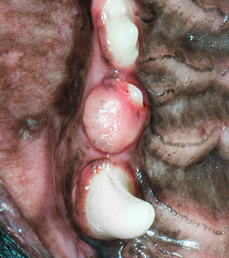
Figure 1. A benign peripheral odontogenic fibroma. (All images courtesy of Dr. Bellows)
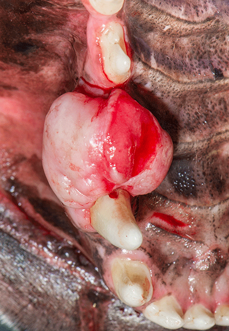
Figure 2. A malignant fibrosarcoma.Set your sights on cytology
The clinical exam can go only so far in identifying oral masses. Touch impression with a cotton applicator swab for cytology or fine-needle aspiration under sedation or anesthesia can help you distinguish between cystic, inflammatory, benign and malignant growths (Figures 3A and 3B). The caveat with cytology is that some neoplasms don't exfoliate, resulting in a nondiagnostic sample or a false negative result. With aspirates, the sample is only as good as where the tip of the needle sampled.
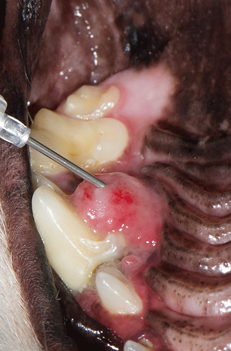
Figure 3A. Obtaining a fine-needle aspiration of a maxillary mass; intraoral radiograph revealed marked tooth resorption.
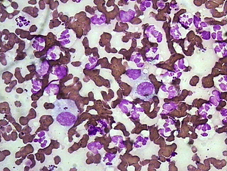
Figure 3B. Cytology consistent with inflammation (mesothelial cells and neutrophils) in the patient from Figure 3A. Carcinomas appear as round or oval cells arranged in sheets or acinar patterns, while sarcomas are spindle-shaped cells. Samples showing at least five cytological signs of malignancy (e.g. high nuclear to cytoplasm ratio, multiple nucleoli, variable nuclear and nucleoli sizes, coarse chromatin, pleomorphism, anisocytosis) are generally diagnostic (Figure 4). False negatives are common. You can generally trust a positive cytology result, but you can't trust a negative one.
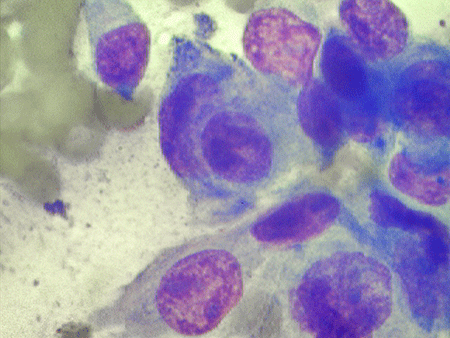
Figure 4. Cytology consistent with malignancy (prominent, different-sized nuclei and nucleoli, nuclear molding, high nuclear-to-cytoplasm ratio); histological diagnosis of squamous cell carcinoma.If you develop confidence in reading real-time cytology, you can save a significant amount of time in treatment staging. If the lesion appears malignant, you can perform excisional surgery with wide margins at the time of the initial procedure. If the lesion does not appear malignant cytologically but does appear so clinically, you have four options: (1) obtain a deep biopsy and wait a week for the histopathology before deciding what to do, (2) refer, (3) remove it with minimal margins with a plan to revisit the lesion if the histopathology is malignant, or (4) excise the mass with wide surgical margins during the initial procedure.
Surgical options for benign oral masses
> Marginal excision removes the lesion visually but may be poorly suited for lesions that are not well demarcated from surrounding healthy tissues. Marginal excision may leave remnants of the tumor in place, resulting in regrowth (Figures 5A-5D).
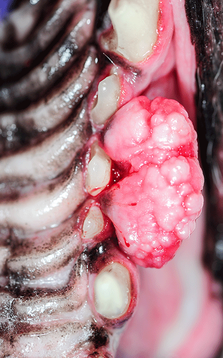
Figure 5A. A right maxillary mass that appears to be emanating from the periodontal ligament spaces rostral and caudal to the second premolar.
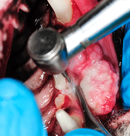
Figure 5B. A marginal excision using a water-cooled bur before extracting the second premolar in the patient from Figure 5A.
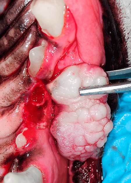
Figure 5C. A caudal pedicle of tissue still attached subgingivally after the second premolar extraction in the patient from Figure 5A.
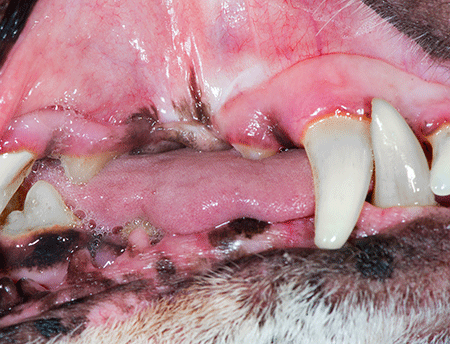
Figure 5D. The healed area in the patient from Figure 5A six months after the operation.> En bloc excision removes the tumor, pseudocapsule and reactive zone, as well as a narrow margin of normal tissue. En bloc excision is usually reserved for malignant or aggressive benign lesions that are not known to extend deep into surrounding tissues (Figures 6A-6C).
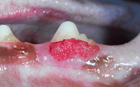
Figure 6A. Mass noted on the buccal attached gingiva.
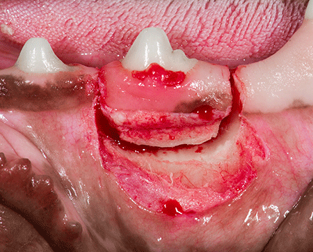
Figure 6B. Gingival flap and en bloc excision of the tooth and mass.
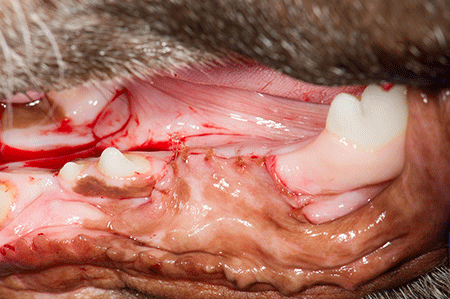
Figure 6C. Sutured surgical site; histopathology showed acanthomatous ameloblastoma completely excised.> Radical resection removes large portions of the maxilla or mandible and is indicated in the treatment of large benign (as well as malignant) tumors that invade the mandible or maxilla (Figures 7A-7D).
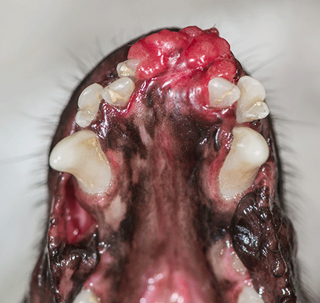
Figure 7A. Large, apparently invasive rostral mandible mass.
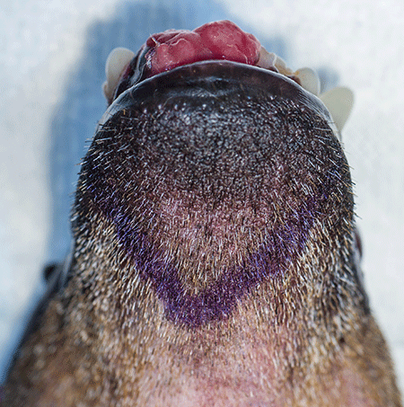
Figure 7B. Incision planning.
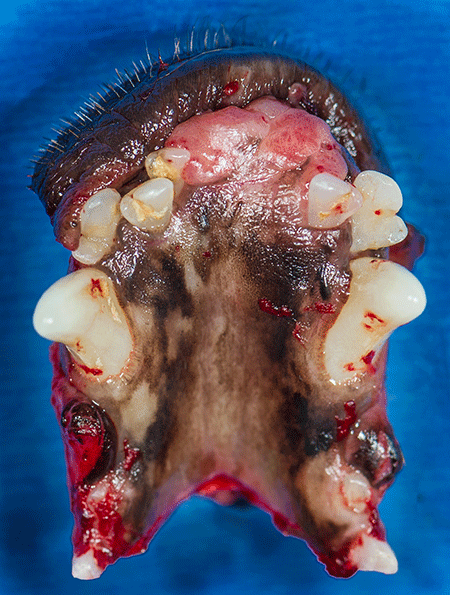
Figure 7C. The radically excised area.
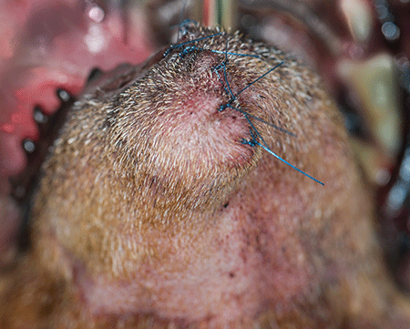
Figure 7D. Sutured surgical site; histologic diagnosis of acanthomatous ameloblastoma.
Commonly diagnosed benign oral masses
> Gingival enlargement is a clinical term referring to the overgrowth or thickening of gingival tissues in the absence of a histological diagnosis (Figure 8). Gingival hyperplasia refers histologically to an abnormal increase in the number of normal cells in a normal arrangement, resulting in gingival enlargement. If you see a boxer, bulldog or cocker spaniel with focal gingival growths, identify the masses as gingival enlargement until you have a histological diagnosis. Patients receiving cyclosporine, anticonvulsants or blood pressure medication are susceptible to developing gingival enlargement. Treatment is usually surgical removal of the abnormal tissues. The condition may recur.
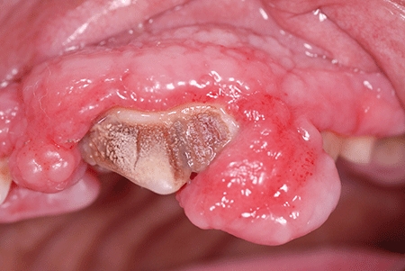
Figure 8. Gingival enlargement.> Eosinophilic granuloma complex affects dogs and cats and causes raised, ulcerated lesions on the tongue, lips and palate (Figures 9A and 9B). These lesions are commonly called indolent or rodent ulcers and are typically slow-growing. In cats, the condition is thought to be connected to flea infestation or food-based allergies. These lesions respond well to corticosteroids, but they may spontaneously regress. Surgical removal is also an option.
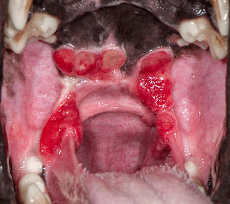
Figure 9A. Eosinophilic granulomas in a dog.
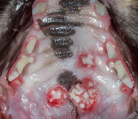
Figure 9B. Eosinophilic granulomas in a cat> Focal fibrous hyperplasia secondary to chronic irritation stemming from subgingival calculus is often seen in brachycephalic breeds including boxers and bulldogs. The clinical appearance is a domelike growth with a smooth mucosal surface of normal coloration (Figure 10). Treatment of these lesions is gingivectomy of the hyperplasia with a scalpel, diode laser or electroscapel. This reestablishes the normal gingival contour and sulcus depth as long as at least 2 mm of attached gingiva remains. Any calculus on the tooth or root surfaces that is covered by the excessive hyperplasia should be scaled and polished away. Daily home plaque control is critical to prevent recurrence.
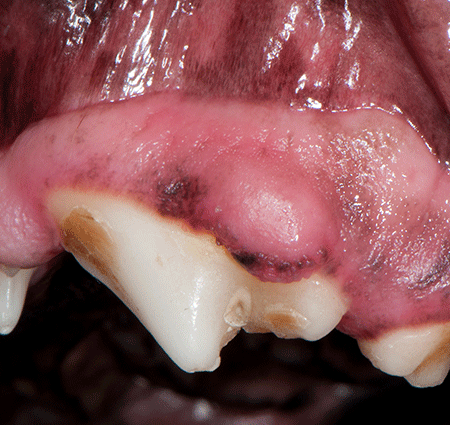
Figure 10. Focal fibrous hyperplasia in a dog.> Peripheral odontogenic fibromas (formerly called fibromatous and ossifying epulides) are quite common in dogs but not in cats (Figure 11A). They can be pedunculated or sessile and may contain osseous material. Complete excision is usually curative (Figure 11B). One study found that 44% of histologically examined epulides in dogs could be classified as focal fibrous hyperplasia, 17% as peripheral odontogenic fibroma and 18% as peripheral ameloblastoma.1
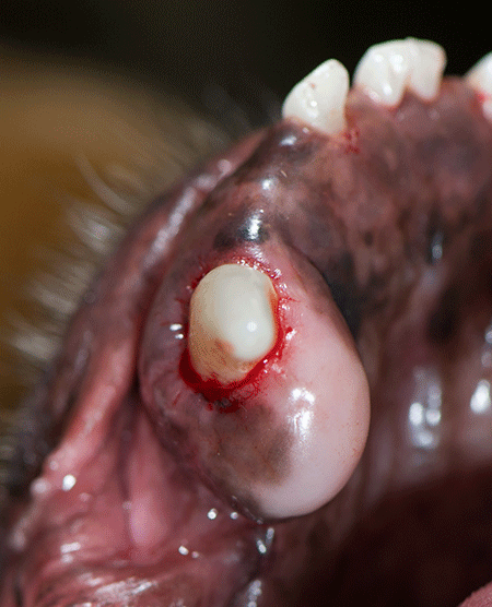
Figure 11A. Peripheral odontogenic fibroma in a dog.
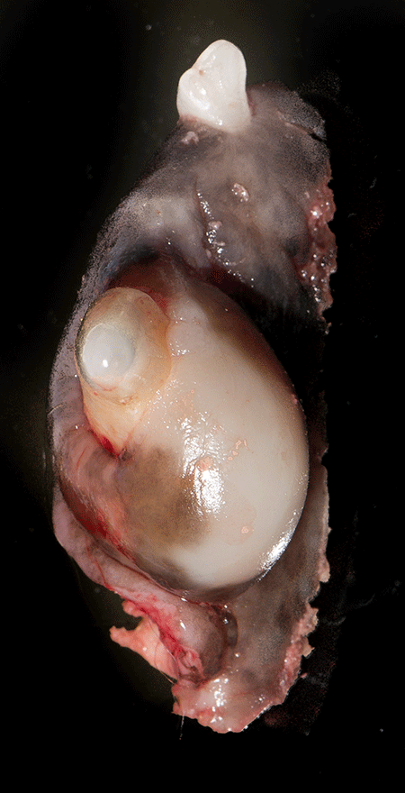
Figure 11B. Excised peripheral odontogenic fibroma.> Acanthomatous ameloblastoma is a benign tumor of odontogenic origin that behaves like a malignant neoplasm but does not metastasize. The most common invasive jaw tumor in dogs, acanthomatous ameloblastoma is locally aggressive and often infiltrates bone. The rostral mandible is the most common location, and the mean age at presentation is 7 to 10 years. This tumor is curable with resection, so 1- to 2-cm margins are ideal whenever functionally possible.
Now that you have the tools to identify and treat benign masses, the next article will explore malignant lesion diagnosis and management.
Reference
1. Verstraete FJ, Ligthelm AJ, Weber A. The histological nature of epiludes in dogs. J Comp Pathol 1992;106(2):169-182.
Further reading
Fiani F, Verstraete FJ, Kass PH, et al. Clinicopathologic characterization of odontogenic tumors and focal fibrous hyperplasia in dogs: 152 cases (1995–2005). J Am Vet Med Assoc 2011;238:495–500.